Identification of a novel oncogene, MMS22L, involved in lung and esophageal carcinogenesis
- Authors:
- Published online on: August 10, 2012 https://doi.org/10.3892/ijo.2012.1589
- Pages: 1285-1296
Abstract
Introduction
Lung cancer is the most common cause of cancer-related death, and the worldwide annual death by lung cancer was estimated to be 1.3 million (1). Esophageal squamous cell carcinoma (ESCC) is one of the most common gastrointestinal tract cancers in Asian countries (2). Although a huge body of knowledge about the biology of lung or esophageal carcinogenesis has been accumulated, the development of novel cancer therapeutics remains inefficient to improve patients with these cancers (3). In fact, in spite of development of various molecular targeted therapies, a limited proportion of patients can receive clinical benefit from them (4).
Through genome-wide gene expression analysis of lung and esophageal cancers, we have isolated a number of oncogenes that were involved in the development and/or progression of cancer (5–41). Among the genes upregulated in these cancers, we focused on MMS22L (methyl methanesulfonate-sensitivity protein 22-like) which is highly expressed in the majority of clinical lung and esophageal cancers. Our original gene expression profile database also revealed that this gene is highly expressed in clinical cervical cancers, but scarcely expressed in normal tissues except testis, suggesting that MMS22L encodes a cancer-testis antigen that can be defined by predominant expression in various types of cancer and undetectable expression in normal tissues except germ cells in testis or ovary (4). Cancer-testis antigens are considered to be good candidate molecular targets for developing new therapeutic strategies for cancers.
Constitutive activation of the NFKB pathway is involved in some forms of cancer such as leukemia, lymphoma, colon cancer and ovarian cancer as well as inflammatory diseases (42–45). The main mechanism of this pathway is reported to be the inactivation of IκB proteins by mutations as well as amplifications and rearrangements of genes encoding the NFKB transcription factor subunits (42–45). However, more commonly it is thought that changes in the upstream pathways that lead to NFKB activation are likely to be aberrantly upregulated in cancer cells (45). Recently some reports suggested that MMS22L-NFKBIL2 interaction could be essential for genomic stability and homologous recombination in immortalized cell lines, suggesting MMS22L to be a new regulator of DNA replication in human cells (46–49). However, no study has indicated critical roles of activation of MMS22L and NFKBIL2 in clinical cancers and investigated their functional importance in carcinogenesis. Here, we report that MMS22L is involved in NFKB pathway in cancer cells through its interaction with NFKBIL2 and might be a promising target for development of novel cancer therapy.
Materials and methods
Cell lines and clinical samples
The 12 human lung-cancer cell lines used for in this study included nine NSCLC cell lines (A549, NCI-H1373, LC319, NCI-H1781, PC-14, NCI-H358, NCI-H2170, NCI-H520 and LU61) and three small-cell lung cancer (SCLC) cell lines (SBC-3, SBC-5 and DMS114). The 9 human esophageal carcinoma cell lines used in this study were as follows: eight SCC cell lines (TE1, TE3, TE8, TE9, TE10, TE12, TE13 and TE15) and one adenocarcinoma (ADC) cell line (TE7). A cervical cancer cell line HeLa was also included in the study. All cells were grown in monolayers in appropriate media supplemented with 10% fetal calf serum (FCS) and were maintained at 37°C in an atmosphere of humidified air with 5% CO2. Human airway epithelial cells, SAEC (Cambrex Bio Science Inc.), were also included in the panel of the cells used in this study. Primary lung and esophageal cancer samples had been obtained earlier with informed consent (5–10). This study and the use of all clinical materials mentioned were approved by individual institutional ethics committees.
Semiquantitative RT-PCR
We prepared appropriate dilutions of each single-stranded cDNA prepared from mRNAs of clinical lung and esophageal cancer samples, taking the level of β-actin (ACTB) expression as a quantitative control. The primer sets for amplification were as follows: ACTB-F (5′-GAGGTGATAGCATTGCTTTCG-3′) and ACTB-R (5′-CAAGTCAGTGTACAGGTAAGC-3′) for ACTB, MMS22L-F (5′-GTCTCACCTTGGACAGATGG-3′) and MMS22L-R (5′-CCAAGGATCCTATTACACAGTTGC-3′) for MMS22L. All reactions involved initial denaturation at 95°C for 5 min followed by 22 (for ACTB) or 30 (for MMS22L) cycles of 95°C for 30 sec, 56°C for 30 sec, and 72°C for 60 sec on a GeneAmp PCR system 9700 (Applied Biosystems).
Northern blot analysis
Human multiple-tissue northern blots (16 normal tissues including heart, brain, placenta, lung, liver, skeletal muscle, kidney, pancreas, spleen, thymus, prostate, testis, ovary, small intestine, colon, leukocyte; BD Biosciences Clontech) were hybridized with a 32P-labeled PCR product of MMS22L. The partial-length cDNA of MMS22L was prepared by RT-PCR using primers MMS22L-F1 (CTGGAAGAGGCAGTTGAAAA) and MMS22L-R1 (ATCGCCCAATATACTGCTCA). Prehybridization, hybridization, and washing were performed according to the supplier’s recommendations. The blots were autoradiographed with intensifying screens at −80°C for 7 days.
Anti-MMS22L antibody
Synthesized peptide with the amino acids sequence of CLGQMGQDEMQRLENDNT [1227–1243] (Cysteine was added to the N-terminal) was inoculated into rabbits; the immune sera were purified on affinity columns according to standard methodology. Affinity-purified anti-MMS22L antibodies were used for western blot as well as immunocytochemical analyses. We confirmed that the antibody was specific to MMS22L on western blots using lysates from cell lines that had been transfected with MMS22L expression vector as well as those from lung and esophageal cancer cell lines that endogenously expressed MMS22L or not.
Western blot analysis
Cells were lysed in lysis buffer; 50 mM Tris-HCl (pH 8.0), 150 mM NaCl, 0.5% NP-40, 0.5% deoxycholate-Na, 0.1% SDS, plus protease inhibitor (Protease Inhibitor Cocktail Set III; Calbiochem). We used ECL western blot analysis system (GE Healthcare Bio-Sciences), as described previously (11).
Immunocytochemical analysis
Cultured cells were washed twice with PBS(-), fixed and rendered permeable in 1:1 acetone: methanol solution for 10 min at −20°C. Prior to the primary antibody reaction, cells were covered with blocking solution [5% bovine serum albumin in PBS(-)] for 10 min to block non-specific antibody binding. After the cells were incubated with a rabbit polyclonal antibody to human MMS22L (generated to synthesized peptide MMS22L; please see above) or a mouse monoclonal antibody to human NFKBIL2 (Abnova), the Alexa Fluor 488-labelled donkey anti-rabbit secondary antibody (Molecular Probes) or Alexa Fluor 594-labbelled donkey anti-mouse secondary antibody (Molecular Probes) was added to detect endogenous MMS22L or NFKBIL2, individually. Nuclei were stained with 4′,6-diamidino-2-phenylindole (DAPI). The antibody-stained cells were viewed with a laser-confocal microscope (TSC SP2 AOBS; Leica Microsystems).
RNA interference assay
Two independent siRNA oligonucleotides against MMS22L were designed using the MMS22L sequences (GenBank accession no: NM198468). Each siRNA (600 pM) was transfected into two NSCLC cell lines, LC319 and A549 or a cervical cancer cell line HeLa using 30 μl of lipofectamine 2,000 (Invitrogen) following the manufacturer’s protocol. The transfected cells were cultured for seven days. Cell numbers and viability were measured by Giemsa staining and 3-(4,5-dimethylthiazol-2-yl)-2,5-diphenyltetrazolium bromide (MTT) assay in triplicate (cell counting kit-8 solution; Dojindo Laboratories). The siRNA sequences used were as follows: control-1 (si-LUC: luciferase gene from Photinus pyralis), 5′-CGUACGCGGAAUACUUCGA-3′; control-2 (CNT: On-TARGETplus siControl non-targeting siRNAs of a pool of four oligosnucleotides: 5′-UGGUUUACAUGUCGACUAA-3′; 5′-UGGUUUACAUGUUUUCUGA-3′; 5′-UGGUUUACAUGUUUUCCUA-3′; and 5′-UGGUUUACAUGUUGUGUGA-3′); siRNA-MMS22L-#1 (si-MMS22L-#1: 5′-CCGCCAAUAUCAUCUCUAAUU-3′); siRNA-MMS22L-#2 (si-MMS22L-#2: 5′-GAACCUGCAAUACAUGGUAUU-3′). Downregulation of endogenous MMS22L expression in the cell lines by siRNAs for MMS22L, but not by controls, was confirmed by semiquantitative RT-PCR and western blot analyses.
Cell growth assay
COS-7 or HEK293 cells that express endogenous MMS22L at a very low level were transfected with mock or MMS22L-expressing vectors (pCAGGSn-3xFlag-MMS22L) using lipofectamine 2,000 transfection reagent (Roche). Transfected cells were incubated in the culture medium containing 0.8 mg/ml neomycin (Geneticin, Invitrogen) for 7 days. Expression of MMS22L as well as viability and colony numbers of cells were evaluated by western blot analysis, and MTT and colony-formation assays at day 7.
Flow cytometric analysis
Cells transfected with siRNA oligonucleotides against MMS22L or control siRNAs were plated at densities of 5x10⁵ per 60-mm dish. Cells were collected in PBS, and fixed in 70% cold ethanol for 30 min. After treatment with 100 μg/ml RNase (Sigma-Aldrich), the cells were stained with 50 μg/ml propidium iodide (Sigma-Aldrich) in PBS. Flow cytometric analysis was done on a Cell Lab Quanta SC (Beckman Coulter) and analyzed by CXP Analysis software (Beckman Coulter). The cells selected from at least 10,000 ungated cells were analyzed for DNA content.
Results
Expression of MMS22L in lung and esophageal cancers
We previously performed genome-wide expression profile analysis of 120 lung cancer cases using microarray consisting of 27,648 cDNAs or ESTs (5–10). Among the genes upregulated in lung and esophageal cancers, we identified MMS22L transcript to be frequently overexpressed in lung and esophageal cancers, and confirmed by semiquantitative RT-PCR experiments its elevated expression in all of eleven clinical lung cancers and in four clinical esophageal cancers, although its expression was not detectable in adjacent normal lung and esophagus tissues (Fig. 1a). We further confirmed by western blot analysis high levels of endogenous MMS22L protein in 11 of 12 lung cancer cell lines and in all of 9 esophageal cancer cell lines using anti-MMS22L antibody (Fig. 1b). Northern blot analysis of 16 normal tissues confirmed that MMS22L was hardly detectable in normal tissues except the testis (Fig. 1c).
Growth effect of MMS22L
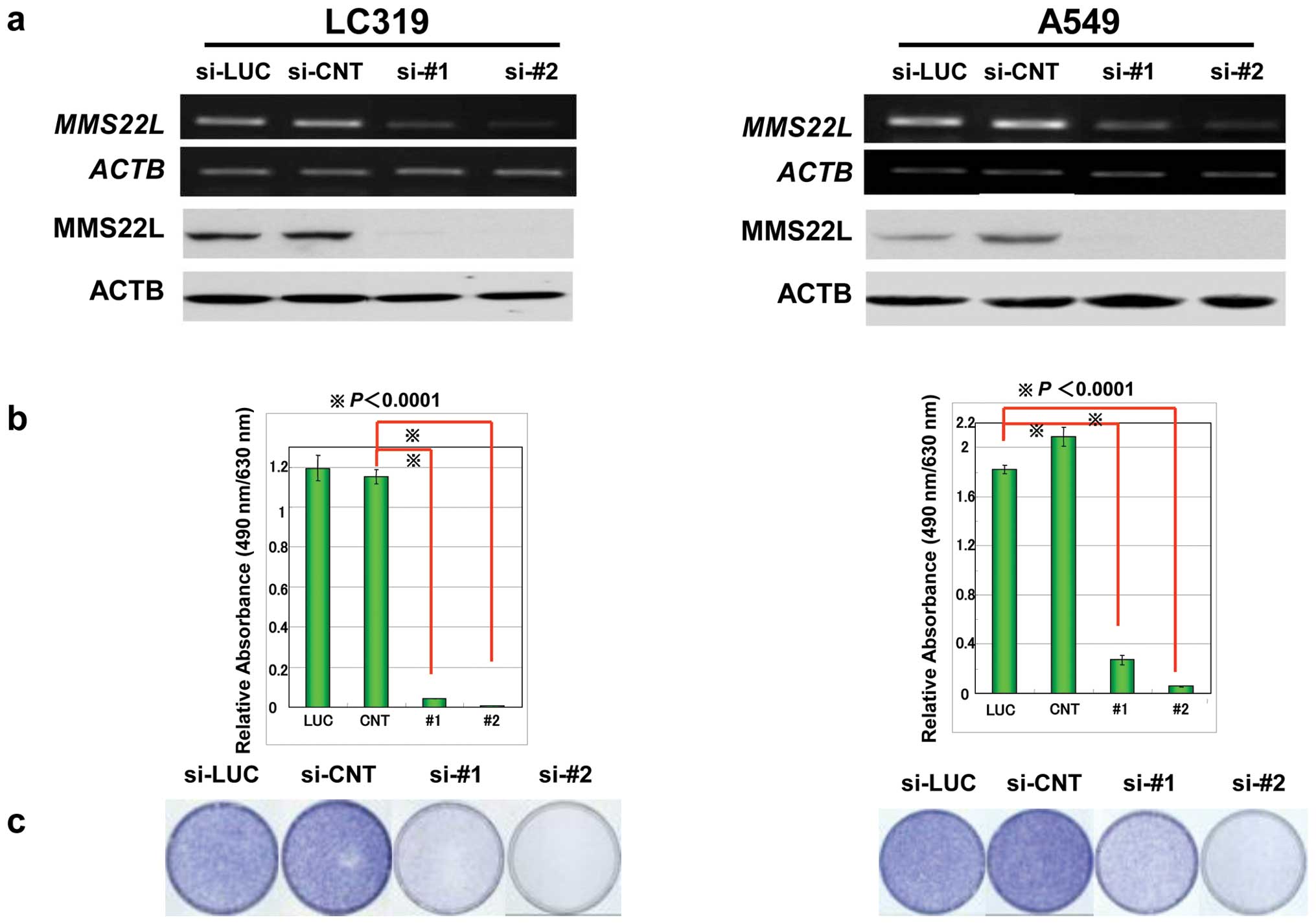
To investigate the relevance of MMS22L to the growth and/or survival of cancer cells, we knocked down the expression of endogenous MMS22L in two lung cancer cell lines, LC319 and A549, by means of the RNAi technique using siRNA oligonucreotide for MMS22L. Semiquantitative RT-PCR experiments detected significant reduction of MMS22L expression in the cells transfected with siRNAs against MMS22L (si-#1 and si-#2), but not in those with control siRNAs (si-LUC and si-CNT) (Fig. 2a). Colony formation and MTT assays clearly demonstrated that the viability of lung cancer cells transfected with two effective siRNAs for MMS22L (si-#1 and si-#2) were reduced in correlation with the reduction of MMS22L expression level, implying essential role of MMS22L in the growth of cancer cells (Fig. 2b and c). Since our original gene expression profile database also revealed its high level of expression in clinical cervical cancers, we also knocked down the expression of MMS22L by siRNAs in a cervical cancer cell line, HeLa, and observed the growth suppressive effect by siRNAs for MMS22L.
To further examine the effect of MMS22L overexpression on the growth of mammalian cells, we transiently transfected plasmid designed to express Flag-tagged MMS22L (pCAGGSn-3xFlag-MMS22L) or mock plasmid into COS-7 or HEK293 cells that expressed endogenous MMS22L at very low level. The significant growth promoting effect was observed in the cells transfected with the MMS22L expressing vector compared to those transfected with the mock vector (Fig. 2d).
NFKBIL2 controls the nuclear localization and stability of MMS22L protein
To investigate the biological function of MMS22L protein, we screened MMS22L-interacting proteins in lung cancer cells using mass spectrometric analysis and identified the interaction between MMS22L and NFKBIL2 [nuclear factor of kappa (NFKB) light polypeptide gene enhancer in B-cells inhibitor-like 2]. Previous reports independently suggested the roles of MMS22L-NFKBIL2 interaction in genomic stability and DNA replication in immortalized cell lines (46–49), however, no study has indicated critical roles of activation of MMS22L and NFKBIL2 in clinical cancers and investigated their functional importance in carcinogenesis. Western blot analysis using cell lines derived from lung cancers and antibodies to MMS22L and NFKBIL2 revealed the co-expression of these two proteins (data not shown), suggesting some functional roles of their interaction in human carcinogenesis. Therefore, we next performed immunofluorescence analysis to determine the subcellular localization of endogenous MMS22L and NFKBIL2 in various cancer cell lines including A549, LC319 and HeLa cells, and found that endogenous MMS22L and NFKBIL2 proteins were mainly co-localized in the nucleus (representative data of HeLa cells was shown in Fig. 3a). To examine the importance of MMS22L-NFKBIL2 interaction in cellar localization of these proteins, we transiently co-expressed exogenous MMS22L and NFKBIL2 proteins using mammalian COS-7 or NIH3T3 cells that expressed these two proteins at very low levels. We found that exogenous MMS22L was mainly located in the cytoplasm and weakly in the nucleus of the cells in which exogenous NFKBIL2 protein was not introduced. However, the nuclear staining of MMS22L was significantly enhanced when both exogenous MMS22L and NFKBIL2 proteins were introduced in the cells (Fig. 3b). On the other hand, exogenous NFKBIL2 was mainly present in the nucleus of cells regardless to the presence or absence of exogenous MMS22L. In addition, we performed western blot analysis using fractionated cytoplasmic and nuclear lysates from COS-7 cells that were introduced exogenous MMS22L and NFKBIL2 proteins. When we transfected both MMS22L-Flag and NFKBIL2-HA expressing vectors, the amounts of nuclear MMS22L was significantly increased, compared with the cells transfected with MMS22L alone (Fig. 3c). Furthermore, we found that knockdown of endogenous MMS22L with siRNA for MMS22L (si-MMS22L) reduced NFKBIL2 protein level in lung cancer LC319 cells and that reduction of NFKBIL2 with si-NFKBIL2 reduced MMS22L levels and significantly suppressed cancer cell growth (Fig. 3d; data not shown). These data suggest that the expression of NFKBIL2 is likely to promote nuclear localization and stability of MMS22L protein, and a complex including these two proteins could coordinately play pivotal roles in cell growth and/or survival.
C-terminal portion of NFKIL2 protein is crucial for binding to MMS22L protein
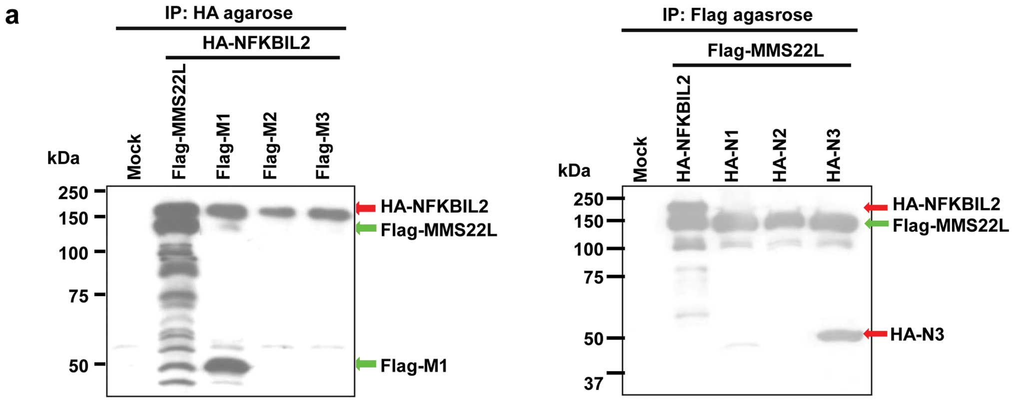
To examine whether the MMS22L-NFKBIL2 protein complex may play important roles in carcinogenesis, we subsequently constructed various plasmids expressing partial MMS22L proteins with Flag tag or partial NFKBIL2 proteins with HA tag, and transfected them into COS-7 cells (data not shown). Immunoprecipitation and western blotting assays using antibodies to Flag- or HA-tags revealed that an N-terminal portion of MMS22L protein (M1; codon 1–414) could bind to a C-terminal region of NFKBIL2 (N3; codon 823–1244) (Fig. 4a). Because immunocytochemical analysis revealed that nuclear localization of MMS22L protein appeared to require the presence of NFKIL2 protein in the nucleus (Fig. 3b), we subsequently investigated which part of NFKBIL2 protein is essential for subcellular localization of MMS22L protein in cultured cells. Plasmids expressing partial proteins of NFKBIL2 were co-transfected with full-length MMS22L expression vector into COS-7 cells. Interestingly, N-terminal (N1; codon 1–450) and central part (N2; codon 403–836) of NFKBIL2 proteins could be localized in the nucleus, while aggregated MMS22L protein was mainly located in the cytoplasm of the same cells (Fig. 4b). It is concordant with the data that these two partial proteins (N1 and N2) could not bind to MMS22L protein as indicated by immunoprecipitation analyses. In contrast, MMS22L protein and C-terminal part of NFKBIL2 protein (N3; codon 823–1244) that could bind to MMS22L protein were mainly localized in the cytoplasm of the cells (Fig. 4b). The data indicate that N-terminal (N1; codon 1–450) and central (N2; codon 403–836) parts of NFKBIL2 are more important for nuclear localization of NFKBIL2, while its C-terminal part (N3; codon 823–1244) is essential for binding to MMS22L.
Dominant negative growth suppressive effect of partial NFKBIL2 protein including MMS22L-binding site
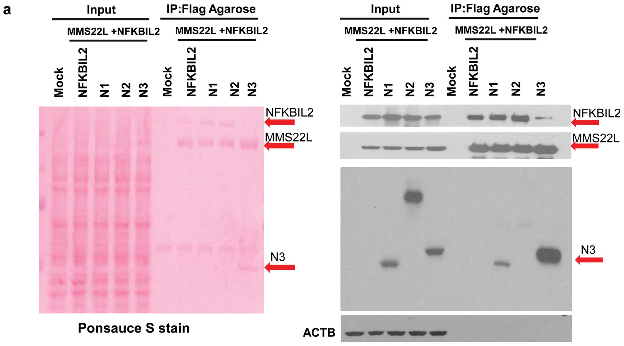
According to the data above, we hypothesized that if nuclear localization of MMS22L protein is important for cancer cells growth, reduction of MMS22L protein in the nucleus by inhibiting the interaction between MMS22L and NFKBIL2 could suppress the cancer cell growth. To examine whether exogenous expression of partial N3 protein can inhibit the MMS22L-NFKBIL2 interaction and cell growth, we co-transfected full-length MMS22L and either of full-/partial-length NFKBIL2 expressing vectors (N1, N2 or N3) into HEK293 cells, and found that the amount of exogenous full-length NFKBIL2 protein that binds to exogenous MMS22L was significantly decreased after introduction of the partial N3 protein, as demonstrated by immunoprecipitation assays, while it was not changed in the cells transfected with N1 or N2 vectors (Fig. 5a). To investigate the functional significance of the interaction between MMS22L and NFKBIL2 for growth of cancer cells, we transfected either of vectors expressing partial NFKBIL2 proteins or mock vectors into two cancer cell lines, HeLa and LC319, which highly expressed both endogenous MMS22L and NFKBIL2 proteins and lung fibroblast CCDlu-19 cells in which MMS22L expression was hardly detectable. Expectedly, exogenous expression of the C-terminal portion of NFKIL2 protein (N3) reduced the levels of MMS22L protein in the nucleus and inhibited the growth of HeLa and LC319 cells as measured by MTT assay, while it did not affect the growth of MMS22L-negative CCDLu-19 cells (Figs. 5b–d). Our findings imply that inhibition of the interaction between the MMS22L and NFKBIL2 protein can suppress the nuclear localization of MMS22L protein, and resulted in the reduction of cancer growth, and that inhibition of the interaction in cancer cells by small molecules might be a potential therapeutic strategy for new cancer treatment.
MMS22L protein acts as an upstream molecule of NFKB pathway
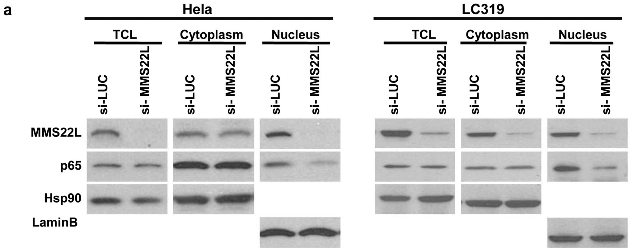
Since NFKIL2 protein was indicated to be involved in the NFKB pathway that plays an essential role in the promotion of cell proliferation and anti-apoptosis (42–45), we examined the expression of NFKB p65/RelA protein in HeLa cells in which both exogenous MMS22L and NFKIL2 were introduced, and found that the level of endogenous p65/RelA protein was elevated compared with those of cells introduced NFKIL2 alone (data not shown). The result suggests that the expression of MMS22L-NFKBIL2 complex may positively regulate the NFKB pathway. Subsequently, we attempted to examine the effect of endogenous MMS22L expression on the NFKB pathway molecules using cytoplasmic and nuclear fraction of HeLa and LC319 cells that were treated with TNF-α. We first confirmed that the level of RelA/p65 was increased in the nucleus of the cells by TNF-α stimulation (data not shown), but that of endogenous RelA/p65 protein was decreased in these cells transfected with siRNA for MMS22L (si-MMS22L) after TNF-α treatment, compared to cells with control siRNA (si-LUC) (Fig. 6a). We then examined the relationship between MMS22L protein and downstream molecules of RelA/p65 such as Bcl-XL and TRAF1/2 that were the anti-apoptosis factors. When we treated the si-LUC-transfected HeLa cells with TNF-α, Bcl-XL and TRAF1 were increased in accordance with the elevation of RelA/p65 (Fig. 6b). However, the elevation of p65, Bcl-XL and TRAF1 were not detected in the TNF-α-stimulated cells transfected with si-MMS22L. The expression level of MMS22L protein showed good correlation with those of p65, Bcl-XL and TRAF1 proteins in lung cancer cell lines (data not shown).
To further examine the effect of MMS22L expression on apoptosis pathway in cancer cells, we cultured cancer cells that were transfected with si-MMS22L under DNA damage condition using DNA-damaging agents (cisplatin/CDDP or 5-fluorouracil/5-FU). After knockdown of MMS22L expression with si-MMS22L in HeLa cells, we treated the cells with CDDP (50 μg/ml) or 5-FU (50 μg/ml) for 48 h and harvested the cells for flow cytometric analysis. The sub G1 population of the cells which were transfected with si-MMS22L was significantly increased compared with those with control siRNA (si-LUC) under DNA damage condition (data not shown). When we exposed the cells that were transfected with si-MMS22L or si-LUC with 20 J of ultraviolet for 48 h, the similar results were observed (data not shown). Western blot analysis using the HeLa cells which were transfected with si-MMS22L or si-LUC, and subsequently treated with CDDP as mentioned above revealed that induction of DNA repair molecules such as ATM, CSB and p53 as well as RelA/p65 and its downstream anti-apoptosis factor Bcl-XL were significantly suppressed in the cells transfected with si-MMS22L compared with those transfected with si-LUC (Fig. 6c). The data suggest that MMS22L can function as an upstream molecule of these anti-apoptosis factors and also affect the induction of some DNA repair pathway molecules (Fig. 6d).
Discussion
Despite the recent development of surgical techniques combined with various treatment modalities such as radiotherapy and chemotherapy, clinical outcome of lung and esophageal cancer patients still remains poor. Therefore, development of new types of anticancer drugs is eagerly awaited. To identify novel target molecules for drug development, we combined genome-wide expression profile analysis of genes that were overexpressed in lung and esophageal cancer cells with high-throughput screening of loss-of-function effects by means of the RNAi technique and tumor tissue microarray analysis (5–41). Through this systematic approach we found MMS22L to be upregulated frequently in clinical lung and esophageal cancer samples, and showed that this gene product plays an indispensable role in the growth and/or survival of cancer cells.
We demonstrated that MMS22L is a putative oncogene and that its nuclear localization and stabilization was enhanced by binding to NFKBIL2. In addition, we revealed that introduction of the C-terminal portion of NFKBIL2 protein into cancer cells could dominant-negatively inhibit the nuclear localization of MMS22L possibly by blocking the MMS22L-NFKBIL2 interaction, and resulted in the suppression of cancer cell growth/ survival. Furthermore, transfection of siRNAs against MMS22L or NFKBIL2 into cancer cells suppressed their expression and the cell growth. Therefore, inhibition of the MMS22L-NFKBIL2 interaction or suppressing MMS22L protein function can be an effective approach for development of novel cancer therapy.
To date, NFKB transcription factors are known to be the key regulators of immune, inflammatory and acute phase responses, and to be involved in the control of cell proliferation and apoptosis (42–45). Activation of NFKB activity and consequent induction of its downstream genes lead to the oncogenesis in mammalian cells. MMS22L protein appeared to act as an upstream molecule of RelA/p65 and be indispensable for induction of anti-apoptosis factors, Bcl-XL or TRAF1. Further studies on the regulation and function of MMS22L protein will contribute to the understanding of molecular mechanism of carcinogenesis through the activation of MMS22L and NFKB pathway.
In cancer chemotherapy, many kinds of DNA damaging agents are being used. The most common approach for targeting the cell cycle is to exploit the effect of DNA-damaging chemotherapeutic agents like 5-FU or CDDP, whose effects are mediated through diverse intracellular targets inducing apoptosis in various cancer cells (50). However, the toxicity of DNA-damaging drugs can be diminished by the activities of several DNA repair pathways as well as anti-apoptotic factors. Therefore, inhibitors of specific DNA repair and/or anti-apoptotic pathways might be promising therapeutic strategy for novel cancer treatments which can improve the efficacy of DNA damage-based cancer therapy (50). Our data suggested the involvement of MMS22L in cellular response to DNA damaging agents. In fact, knockdown of MMS22L expression also enhanced the apoptosis of cancer cells that were exposed to DNA-damaging agents including 5-FU and CDDP probably due to inhibition of induction of DNA repair molecules such as ATM, CSB and p53 as well as RelA/p65 and its downstream anti-apoptosis factor Bcl-XL. The combined data of our experiments suggest that MMS22L might function as an upstream molecule of these anti-apoptosis factors and DNA-repair molecules and that targeting MMS22L could have a significant advantage in avoiding the resistance of cancer cells to anticancer treatments, although the detailed function of MMS22L in drug response of the cells and in carcinogenesis remains to be elucidated.
In summary, our data indicate that MMS22L is involved in NFKB pathway in cancer cells through its interaction with NFKBIL2 and that it might be a promising candidate target for developing highly specific anticancer drugs with minimal risk of adverse effects.
Acknowledgements
This study was supported in part by Grant-in-Aid for Scientific Research (B) and Grant-in-Aid for Scientific Research on Innovative Areas from The Japan Society for the Promotion of Science to Y.D. Y.D. is a member of Shiga Cancer Treatment Project supported by Shiga Prefecture (Japan).
References
|
Jemal A, Siegel R, Ward E, Hao Y, Xu J and Thun MJ: Cancer statistics, 2009. CA Cancer J Clin. 59:225–249. 2009. View Article : Google Scholar | |
|
Shimada H, Nabeya Y, Okazumi S, et al: Prediction of survival with squamous cell carcinoma antigen in patients with resectable esophageal squamous cell carcinoma. Surgery. 133:486–494. 2003. View Article : Google Scholar : PubMed/NCBI | |
|
Berwick M and Schantz S: Chemoprevention of aerodigestive cancer. Cancer Metastasis Rev. 16:329–347. 1997. View Article : Google Scholar : PubMed/NCBI | |
|
Daigo Y and Nakamura Y: From cancer genomics to thoracic oncology: discovery of new biomarkers and therapeutic targets for lung and esophageal carcinoma. Gen Thorac Cardiovasc Surg. 56:43–53. 2008. View Article : Google Scholar : PubMed/NCBI | |
|
Kikuchi T, Daigo Y, Katagiri T, et al: Expression profiles of non-small cell lung cancers on cDNA microarrays: identification of genes for prediction of lymph-node metastasis and sensitivity to anti-cancer drugs. Oncogene. 22:2192–205. 2003. View Article : Google Scholar : PubMed/NCBI | |
|
Kakiuchi S, Daigo Y, Tsunoda T, Yano S, Sone S and Nakamura Y: Genome-wide analysis of organ-preferential metastasis of human small cell lung cancer in mice. Mol Cancer Res. 1:485–499. 2003.PubMed/NCBI | |
|
Kakiuchi S, Daigo Y, Ishikawa N, et al: Prediction of sensitivity of advanced non-small cell lung cancers to gefitinib (Iressa, ZD1839). Hum Mol Genet. 13:3029–43. 2004. View Article : Google Scholar : PubMed/NCBI | |
|
Kikuchi T, Daigo Y, Ishikawa N, et al: Expression profiles of metastatic brain tumor from lung adenocarcinomas on cDNA microarray. Int J Oncol. 28:799–805. 2006.PubMed/NCBI | |
|
Taniwaki M, Daigo Y, Ishikawa N, et al: Gene expression profiles of small-cell lung cancers: molecular signatures of lung cancer. Int J Oncol. 29:567–575. 2006.PubMed/NCBI | |
|
Yamabuki T, Daigo Y, Kato T, et al: Genome-wide gene expression profile analysis of esophageal squamous cell carcinomas. Int J Oncol. 28:1375–1384. 2006.PubMed/NCBI | |
|
Suzuki C, Daigo Y, Kikuchi T, Katagiri T and Nakamura Y: Identification of COX17 as a therapeutic target for non-small cell lung cancer. Cancer Res. 63:7038–7041. 2003.PubMed/NCBI | |
|
Kato T, Daigo Y, Hayama S, et al: A novel human tRNA-dihydrouridine synthase involved in pulmonary carcinogenesis. Cancer Res. 65:5638–5646. 2005. View Article : Google Scholar : PubMed/NCBI | |
|
Furukawa C, Daigo Y, Ishikawa N, et al: Plakophilin 3 oncogene as prognostic marker and therapeutic target for lung cancer. Cancer Res. 65:7102–7110. 2005. View Article : Google Scholar : PubMed/NCBI | |
|
Suzuki C, Daigo Y, Ishikawa N, et al: ANLN plays a critical role in human lung carcinogenesis through the activation of RHOA and by involvement in the phosphoinositide 3-kinase/AKT pathway. Cancer Res. 65:11314–11325. 2005. View Article : Google Scholar : PubMed/NCBI | |
|
Ishikawa N, Daigo Y, Takano A, et al: Characterization of SEZ6L2 cell-surface protein as a novel prognostic marker for lung cancer. Cancer Sci. 97:737–745. 2006. View Article : Google Scholar : PubMed/NCBI | |
|
Takahashi K, Furukawa C, Takano A, et al: The neuromedin u-growth hormone secretagogue receptor 1b/neurotensin receptor 1 oncogenic signaling pathway as a therapeutic target for lung cancer. Cancer Res. 66:9408–9419. 2006. View Article : Google Scholar : PubMed/NCBI | |
|
Hayama S, Daigo Y, Kato T, et al: Activation of CDCA1-KNTC2, members of centromere protein complex, involved in pulmonary carcinogenesis. Cancer Res. 66:10339–10348. 2006. View Article : Google Scholar : PubMed/NCBI | |
|
Kato T, Hayama S, Yamabuki T, et al: Increased expression of IGF-II mRNA-binding protein 1 is associated with the tumor progression in patients with lung cancer. Clin Cancer Res. 13:434–442. 2007. View Article : Google Scholar : PubMed/NCBI | |
|
Suzuki C, Takahashi K, Hayama S, et al: Identification of Myc-associated protein with JmjC domain as a novel therapeutic target oncogene for lung cancer. Mol Cancer Ther. 6:542–551. 2007. View Article : Google Scholar : PubMed/NCBI | |
|
Hayama S, Daigo Y, Yamabuki T, et al: Phosphorylation and activation of cell division cycle associated 8 by aurora kinase B plays a significant role in human lung carcinogenesis. Cancer Res. 67:4113–4122. 2007. View Article : Google Scholar : PubMed/NCBI | |
|
Taniwaki M, Takano A, Ishikawa N, et al: Activation of KIF4A as a prognostic biomarker and therapeutic target for lung cancer. Clin Cancer Res. 13:6624–6631. 2007. View Article : Google Scholar : PubMed/NCBI | |
|
Mano Y, Takahashi K, Ishikawa N, et al: Fibroblast growth factor receptor 1 oncogene partner as a novel prognostic biomarker and therapeutic target for lung cancer. Cancer Sci. 98:1902–1913. 2007. View Article : Google Scholar : PubMed/NCBI | |
|
Kato T, Sato N, Hayama S, et al: Activation of holliday junction recognizing protein involved in the chromosomal stability and immortality of cancer cells. Cancer Res. 67:8544–8553. 2007. View Article : Google Scholar : PubMed/NCBI | |
|
Kato T, Sato N, Takano A, et al: Activation of placenta specific transcription factor distal-less homeobox 5 predicts clinical outcome in primary lung cancer patients. Clin Cancer Res. 14:2363–2370. 2008. View Article : Google Scholar : PubMed/NCBI | |
|
Dunleavy EM, Roche D, Tagami H, et al: HJURP is a cell-cycle-dependent maintenance and deposition factor of CENP-A at centromeres. Cell. 137:485–497. 2009. View Article : Google Scholar : PubMed/NCBI | |
|
Hirata D, Yamabuki T, Miki D, et al: Involvement of epithelial cell transforming sequence-2 oncoantigen in lung and esophageal cancer progression. Clin Cancer Res. 15:256–266. 2009. View Article : Google Scholar : PubMed/NCBI | |
|
Sato N, Koinuma J, Fujita M, et al: Activation of WD repeat and high-mobility group box DNA binding protein 1 in pulmonary and esophageal carcinogenesis. Clin Cancer Res. 16:226–239. 2010. View Article : Google Scholar : PubMed/NCBI | |
|
Sato N, Koinuma J, Ito T, et al: Activation of an oncogenic TBC1D7 (TBC1 domain family, member 7) protein in pulmonary carcinogenesis. Genes Chromosomes Cancer. 49:353–367. 2010.PubMed/NCBI | |
|
Nguyen MH, Koinuma J, Ueda K, et al: Phosphorylation and activation of cell division cycle associated 5 by mitogen-activated protein kinase play a crucial role in human lung carcinogenesis. Cancer Res. 70:5337–5347. 2010. View Article : Google Scholar : PubMed/NCBI | |
|
Ishikawa N, Daigo Y, Yasui W, et al: ADAM8 as a novel serological and histochemical marker for lung cancer. Clin Cancer Res. 10:8363–8370. 2004. View Article : Google Scholar : PubMed/NCBI | |
|
Ishikawa N, Daigo Y, Takano A, et al: Increases of amphiregulin and transforming growth factor-alpha in serum as predictors of poor response to gefitinib among patients with advanced non-small cell lung cancers. Cancer Res. 65:9176–9184. 2005. View Article : Google Scholar : PubMed/NCBI | |
|
Yamabuki T, Takano A, Hayama S, et al: Dickkopf-1 as a novel serologic and prognostic biomarker for lung and esophageal carcinomas. Cancer Res. 67:2517–2525. 2007. View Article : Google Scholar : PubMed/NCBI | |
|
Ishikawa N, Takano A, Yasui W, et al: Cancer-testis antigen lymphocyte antigen 6 complex locus K is a serologic biomarker and a therapeutic target for lung and esophageal carcinomas. Cancer Res. 67:11601–11611. 2007. View Article : Google Scholar | |
|
Takano A, Ishikawa N, Nishino R, et al: Identification of nectin-4 oncoprotein as a diagnostic and therapeutic target for lung cancer. Cancer Res. 69:6694–6703. 2009. View Article : Google Scholar : PubMed/NCBI | |
|
Sato N, Yamabuki T, Takano A, et al: Wnt inhibitor Dickkopf-1 as a target for passive cancer immunotherapy. Cancer Res. 70:5326–5336. 2010. View Article : Google Scholar : PubMed/NCBI | |
|
Suda T, Tsunoda T, Daigo Y, Nakamura Y and Tahara H: Identification of human leukocyte antigen-A24-restricted epitope peptides derived from gene products upregulated in lung and esophageal cancers as novel targets for immunotherapy. Cancer Sci. 98:1803–1808. 2007. View Article : Google Scholar : PubMed/NCBI | |
|
Mizukami Y, Kono K, Daigo Y, et al: Detection of novel cancer-testis antigen-specific T-cell responses in TIL, regional lymph nodes, and PBL in patients with esophageal squamous cell carcinoma. Cancer Sci. 99:1448–1454. 2008. View Article : Google Scholar : PubMed/NCBI | |
|
Harao M, Hirata S, Irie A, et al: HLA-A2-restricted CTL epitopes of a novel lung cancer-associated cancer testis antigen, cell division cycle associated 1, can induce tumor-reactive CTL. Int J Cancer. 123:2616–2625. 2008. View Article : Google Scholar : PubMed/NCBI | |
|
Kono K, Mizukami Y, Daigo Y, et al: Vaccination with multiple peptides derived from novel cancer-testis antigens can induce specific T-cell responses and clinical responses in advanced esophageal cancer. Cancer Sci. 100:1502–1509. 2009. View Article : Google Scholar : PubMed/NCBI | |
|
Yokomine K, Senju S, Nakatsura T, et al: The forkhead box M1 transcription factor, as a candidate of target for anti-cancer immunotherapy. Int J Cancer. 126:2153–2163. 2010.PubMed/NCBI | |
|
Tomita Y, Imai K, Senju S, et al: A novel tumor-associated antigen, cell division cycle 45-like can induce cytotoxic T-lymphocytes reactive to tumor cells. Cancer Sci. 102:697–705. 2011. View Article : Google Scholar : PubMed/NCBI | |
|
Rayet B and Gelinas C: Aberrant rel/nfkb genes and activity in human cancer. Oncogene. 18:6938–6947. 1999. View Article : Google Scholar : PubMed/NCBI | |
|
Tergaonkar V: NFκB pathway: A good signaling paradigm and therapeutic target. Int J Biochem Cell Biol. 38:1647–1653. 2006. | |
|
Yamamoto Y and Gaynor RB: Therapeutic potential of inhibition of the NF-kappaB pathway in the treatment of inflammation and cancer. J Clin Invest. 107:135–142. 2001. View Article : Google Scholar : PubMed/NCBI | |
|
Kim HJ, Hawke N and Baldwin AS: NF-κB and IKK as therapeutic targets in cancer. Cell Death Differ. 13:738–747. 2006. | |
|
O’Donnell L, Panier S, Wildenhain J, et al: The MMS22L-TONSL complex mediates recovery from replication stress and homologous recombination. Mol Cell. 40:619–631. 2010.PubMed/NCBI | |
|
Duro E, Lundin C, Ask K, et al: Identification of the MMS22L-TONSL complex that promotes homologous recombination. Mol Cell. 40:632–644. 2010. View Article : Google Scholar : PubMed/NCBI | |
|
Brenda C, O’Connell L, Adamson B, et al: A genome-wide camptothecin sensitivity screen identifies a mammalian MMS22L-NFKBIL2 complex required for genomic stability. Mol Cell. 40:645–657. 2010. View Article : Google Scholar : PubMed/NCBI | |
|
Piwko W, Olma MH, Held M, et al: RNAi-based screening identifies the Mms22L-Nfkbil2 complex as a novel regulator of DNA replication in human cells. EMBO J. 29:4210–4222. 2010. View Article : Google Scholar : PubMed/NCBI | |
|
Lee BJ, Chon KM, Kim YS, et al: Effects of cisplatin, 5-fluorouracil, and radiation on cell cycle regulation and apoptosis. Chemotherapy. 51:103–110. 2005. View Article : Google Scholar : PubMed/NCBI |


